
Tests
What is a cardiac MRI scan and when is it needed?
What a cardiac MRI scan shows that an echocardiogram and a CT cannot, when it is genuinely needed, and what happens during the scan itself.
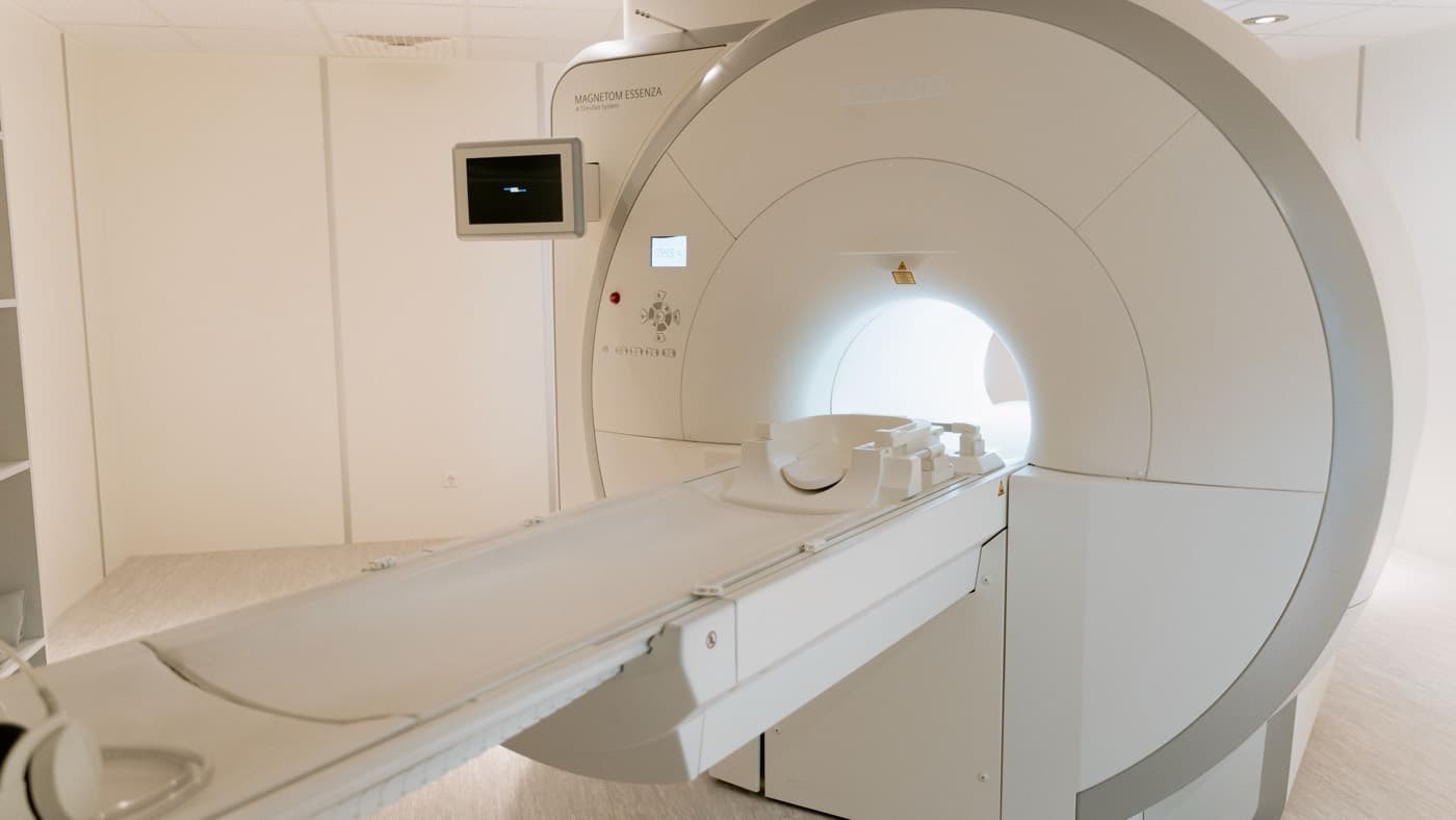
A cardiac MRI scan uses a strong magnetic field and radio waves, rather than radiation, to produce extremely detailed images of the heart muscle itself. It is the most informative look at the structure and texture of the heart that we currently have.
That last point is what makes it different from the other scans. An echocardiogram shows how well the heart is pumping. A cardiac CT looks at the arteries supplying it. A cardiac MRI answers a third question, which is what the heart muscle is actually made of and whether any of it has been damaged, inflamed or replaced.
What It Shows That Other Tests Cannot
Most cardiac tests measure function. MRI characterises tissue, and that is the distinction worth understanding.
By giving a contrast agent called gadolinium and imaging the heart a few minutes later, we can see where that contrast lingers in the muscle. Healthy heart muscle clears it. Scarred or inflamed muscle does not. The pattern of what lights up, and crucially where it sits within the wall of the heart, tells us a great deal about the cause.
Scar in one particular distribution points to a previous heart attack, sometimes one nobody knew had happened. Scar in a different pattern points instead to inflammation of the heart muscle, or to one of the cardiomyopathies, or to an infiltrative condition where an abnormal substance is deposited in the muscle. Those look similar on an echocardiogram and are managed completely differently.
Cardiac MRI also measures the volume of the chambers and the ejection fraction more accurately and more reproducibly than ultrasound, which matters when a treatment decision hangs on whether a number has genuinely changed.
How It Compares With an Echo and a CT
Patients often ask which scan is the best one, and the honest answer is that they are not competing. They answer different questions.
An echocardiogram is an ultrasound. It is quick, involves no radiation and no injection, and it is excellent at assessing the valves and at giving a working measure of how the heart is pumping. It is almost always the right first test, and for many people it is the only one needed.
A cardiac CT is fast and looks at the coronary arteries in detail, which makes it the test of choice when the question is whether those arteries are narrowed. It uses radiation and an iodine based contrast dye.
A cardiac MRI takes longer than either and uses no radiation, and it is the one that characterises the muscle itself.
Put simply, if the question is how well the heart is working, an echocardiogram usually answers it. If the question is whether the arteries are narrowed, CT usually answers it. If the question is why this heart is abnormal in the first place, MRI is frequently the study that provides the answer.
None of the three replaces the others and it is common to need more than one. What should not happen is a scan being chosen because it was available rather than because it answered your question.
When It Is Actually Needed
It is a specialist test rather than a routine one, and these are the situations where it earns its place.
- Unexplained heart failure or a weakened heart. When the pumping function is reduced and the reason is not obvious, MRI is often what identifies the cause. That changes treatment, and sometimes it changes the advice given to a patient's family.
- Suspected cardiomyopathy. For distinguishing between the different types, and for assessing risk, MRI does things no other test does. How cardiomyopathy affects the heart depends heavily on which type it is.
- Thickened heart muscle. An echocardiogram can tell you the wall is thick. MRI is far better at telling you why, which might be long standing high blood pressure, an athlete's heart, hypertrophic cardiomyopathy or an infiltrative disease.
- Suspected inflammation of the heart muscle, particularly in a younger person with chest pain and abnormal blood tests but normal coronary arteries.
- Before certain rhythm procedures. Scar in the heart muscle is often the substrate for heart rhythm problems, so knowing where it is can inform both risk and planning.
- Assessing blood supply without radiation. Stress perfusion MRI can show whether areas of muscle are getting enough blood during stress, which is an alternative to nuclear imaging for suspected coronary artery disease.
What Happens on the Day
An MRI scan is painless, but it asks more of you than most tests, and knowing that in advance helps.
You lie on a bed that moves into a tube shaped scanner. It is noisy, so you will be given headphones or ear protection. Electrodes on the chest let the scanner synchronise its images with your heartbeat, and you will be asked to hold your breath for perhaps 10 to 15 seconds at a time, repeatedly. Most cardiac MRI studies take between 30 and 60 minutes, which is considerably longer than a CT.
If contrast is needed, a cannula goes into a vein in your arm. Kidney function is checked first.
Two things genuinely matter beforehand. If you are claustrophobic, say so early, because it can be planned for rather than discovered halfway through. And if you have a pacemaker, a defibrillator or any metal implant, that must be declared. Many modern devices are MRI conditional and scanning is possible with the right protocol, but this needs establishing in advance rather than on the day.
Why the Interpretation Matters More Than the Scan
Cardiac MRI produces a large amount of data, and the value sits in reading it in context. The same pattern of scar means one thing in a 30 year old with palpitations and something quite different in a 70 year old with breathlessness and known coronary disease.
This is the area Dr Jogiya trained in. His PhD was in cardiac MRI at King's College London, funded by a British Heart Foundation fellowship, and he holds SCMR level 3 from the Society for Cardiovascular Magnetic Resonance. His published research is largely in stress perfusion cardiac MRI and its validation against invasive measurements of coronary disease, and it has been cited more than a thousand times.
That matters because it shapes how the test is used. Cardiac MRI is often more useful than repeating an echocardiogram when the question has shifted from how badly the heart is affected to why it is affected at all, because a second ultrasound usually confirms what the first one showed rather than explaining it. Where the question really is about severity or valve function over time, a repeat echo is the right and much simpler answer.
Only a small proportion of patients need a cardiac MRI. It is not a screening test and it is not a general heart check. But where the diagnosis genuinely hangs on what the muscle looks like, it is often the study that settles the question after everything else has been inconclusive.
Conclusion
A cardiac MRI scan is a detailed, radiation free study of the heart muscle that answers questions other tests cannot, particularly about scarring, inflammation and the cause of a weakened or thickened heart. It takes longer than a CT, involves lying still in a noisy scanner with repeated breath holds, and needs planning if you are claustrophobic or have an implanted device.
It is a targeted test rather than a routine one, and the useful version of the question is not whether you can have one but whether it will change what happens next. Alongside CT scans of the heart and echocardiography it forms a set, and choosing correctly between them is most of the skill.
If you have been recommended a cardiac MRI, if you have a report you would like explained, or you have unexplained breathlessness or a diagnosis of living with heart failure that has never been fully accounted for, you can contact me, Dr Roy Jogiya, at Kingston Cardiologist to arrange a proper cardiac assessment in Kingston upon Thames or central London. Appointments are available in person and virtually, with full diagnostic support including advanced cardiac imaging.
Keep reading