
Tests
Echocardiogram Test Kingston
Book an echocardiogram in Kingston with consultant-led review, clear results and prompt access to specialist assessment.
If you're searching for an echocardiogram test in Kingston, the short answer is that Kingston Cardiologist offers prompt access to specialist assessment, and investigations can often be coordinated alongside or following consultation. An echocardiogram is an ultrasound scan of the heart that uses harmless sound waves to produce a live, moving picture of how your heart is working. It's painless, safe, involves no radiation, and takes around 30 to 45 minutes. It is the usual first-line test for answering structural questions about the heart.
For local patients, the practical question often isn't what an echo is, it's how to arrange one, who will perform it, and how the result fits into a wider assessment of their symptoms. At Kingston Cardiologist, an echocardiogram is performed and interpreted by a consultant cardiologist rather than reported separately later, and investigations can often be coordinated alongside or following consultation, with the findings discussed face to face.
What an Echocardiogram Test Actually Shows
The heart is a mechanical pump as well as an electrical one, and an echo is how we see the mechanics. The scan shows the size and shape of each of the four chambers, the thickness and movement of the heart muscle, how strongly the main pumping chamber (the left ventricle) is contracting, and how each of the four valves is opening and closing in real time. Using a Doppler element, it also measures the speed and direction of blood flow, which lets us calculate pressures inside the heart without any need for invasive testing.
That breadth is what makes the echo so valuable. A single 30-minute scan can confirm or rule out heart valve disease, heart failure, cardiomyopathy, the structural consequences of high blood pressure, congenital differences picked up later in life, and the after-effects of a previous heart attack. Few tests offer this much information for so little inconvenience to the patient.
Why People in Kingston Get Referred for an Echo
The conditions and symptoms that most commonly prompt an echocardiogram referral are predictable, and the threshold for arranging one is generally low. The test is safe and informative, so there is rarely a good reason not to do one when there is a real cardiac question to answer. Common reasons for referral include:
Unexplained breathlessness, particularly during light activity or when lying flat
A heart murmur picked up on examination
Persistent or new chest pain
Palpitations or known rhythm disorders such as atrial fibrillation
Ankle or leg swelling that might point to fluid retention
Dizziness or fainting episodes
A family history of cardiomyopathy, congenital heart disease or sudden cardiac death
Follow-up after a previous heart attack
Baseline monitoring for patients on chemotherapy drugs that can affect the heart
Pre-participation assessment for athletes returning to high-intensity training
A growing number of patients are also referred after an irregular reading from a smartwatch or fitness tracker. An echocardiogram adds the structural picture that wearable data cannot provide, so the two together build a much fuller assessment than either on its own.
What an Echocardiogram Test Appointment Looks Like in Kingston
At Kingston Cardiologist, an echocardiogram test appointment is built around a consultation, not around the scan in isolation. A typical visit unfolds in roughly this sequence.
You'll begin with a detailed history, where we talk through your symptoms, your medical background, your family history and anything else relevant. This usually takes around 15 to 20 minutes and is the part of the appointment that most often shapes the rest of the assessment.
A physical examination follows, including listening to your heart and lungs, checking your pulse and blood pressure, and looking for signs of fluid retention or other clinical findings.
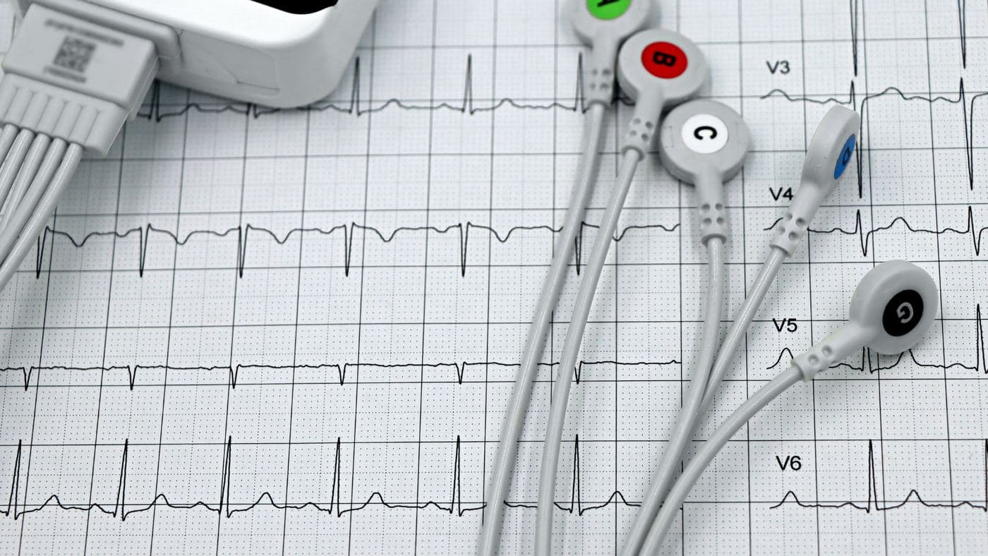
An ECG can often be carried out alongside the consultation. It's a five-minute test that records the electrical activity of your heart and complements the structural information from the echo.
The echocardiogram itself is performed next. You'll be asked to remove clothing from the waist up, lie on your left side on a couch, and a small amount of clear gel is applied to your chest. A handheld probe is then moved gently across different parts of your chest, capturing images from multiple angles. You'll often hear the rhythmic whooshing sound of blood flow being measured. The whole scan takes between 30 and 45 minutes. There's no pain, no radiation, no needles and no preparation needed beforehand.
Finally, where the scan is carried out at the consultation, the findings are discussed with you face to face before you leave, with a clear plan agreed and any onward investigations explained. Having the findings explained in person is what turns a set of measurements into something you can act on.
Why Consultant-Led Echocardiography Matters
The quality of an echocardiogram depends on the equipment, but it depends even more on the experience of the person performing and interpreting the images. A consultant-led echocardiogram has a practical advantage over a separately reported scan, because the cardiologist running the scan can adapt it in real time to the clinical question. If something interesting appears in a standard view, more time can be spent on it. If a specific concern needs answering, additional views can be added on the spot.
Bringing the history, examination, ECG and echocardiogram together in one place is what makes sense of the findings, and it is not unusual for the scan to confirm a working diagnosis or to prompt a rethink. That is the reason consultations are run with the equipment ready on site rather than referring scans out to a separate department.
The other practical advantage is straightforward. Where investigations can be coordinated alongside the consultation, there is no need to attend two or three separate appointments to build a complete picture. For patients balancing work, family and the worry that comes with cardiac symptoms, that makes a meaningful difference.
How an Echocardiogram Test Compares to Other Heart Tests
Patients often ask how an echocardiogram differs from other heart investigations they've heard of.
ECG (electrocardiogram). An ECG records electrical activity, while an echo shows structure and function. The two answer different questions and are usually best done together rather than as alternatives.
Cardiac MRI. A cardiac MRI provides exceptionally detailed images of the heart muscle and is particularly useful for assessing scarring, inflammation and certain cardiomyopathies. It's a more specialised test and takes around an hour, but it isn't usually a first-line investigation.
CT coronary angiogram. A CT angiogram looks specifically at the coronary arteries that supply the heart muscle, which an echo doesn't directly visualise. The two tests are complementary in the workup of chest pain.
Invasive coronary angiogram. This is reserved for patients in whom non-invasive testing has raised significant concern about coronary artery disease, and it carries the option of treating a blockage in the same sitting.
An echocardiogram is usually the most useful first structural test in a new cardiology assessment. Most questions about the structure of the heart can be settled with a standard echo, and where they cannot, a more specialised scan can be arranged when the clinical picture warrants it.
Specialised Types of Echocardiogram
When patients are referred for an echo, what they typically have is a standard transthoracic echocardiogram (TTE). This is the scan described above and answers most clinical questions. There are also more specialised forms.
A stress echocardiogram combines the scan with exercise or a medication that stresses the heart, so we can see how the heart muscle behaves under load. This is useful for suspected coronary artery disease or borderline valve problems.
A transoesophageal echocardiogram (TOE) uses a small probe passed down the throat into the oesophagus under sedation. Because the oesophagus sits directly behind the heart, the images are exceptionally clear, and this scan is used for detailed valve assessment or to look for clots before certain procedures.
A contrast echocardiogram involves a small intravenous injection of microbubbles, which makes the heart chambers show up more clearly. This helps in patients whose body shape or lung tissue makes a standard scan harder to interpret.
The simplest test that will answer the question is usually the right one, and that is normally a standard TTE. A specialised type is added only when there is a specific reason.
Common Worries About the Test
A few questions come up regularly in clinic and are worth addressing directly.
Is it safe? Yes, completely. There's no radiation, no needles for a standard scan, and no known harmful effects. It's safe in pregnancy and safe to repeat as often as needed.
Will it pick up a heart murmur? An echo doesn't directly record sound the way a stethoscope does, but it will show the underlying cause of any murmur, which is far more useful clinically.
Can a normal scan miss heart problems? Sometimes. An echo is excellent for structure and function but doesn't directly visualise the coronary arteries. If your symptoms suggest coronary disease, additional tests may be needed.
Does an abnormal finding always mean something serious? No. Many findings are minor or normal variants. Mild valve leaks, for example, are extremely common and usually need no treatment at all. The interpretation in context is what matters.
When to Arrange an Echocardiogram
Some symptoms need urgent attention rather than a routine appointment. Chest pain or pressure that is severe, lasts more than 15 minutes or does not settle with rest, sudden severe breathlessness, or collapsing or blacking out all mean calling 999. If symptoms are worrying but not an emergency, contact NHS 111 or ask your GP for an urgent appointment. Once anything urgent has been excluded, breathlessness that is new or progressive, ankle or leg swelling that does not settle, chest discomfort brought on by exertion, frequent palpitations, or any new symptom in someone with a strong family history of heart disease are all good reasons to arrange a proper cardiology review.
Symptoms are often explained away for weeks or months as stress, indigestion or simply getting older. The earlier they are properly evaluated, the more straightforward the answers tend to be, and the more options are available if treatment is ever needed. An echocardiogram, combined with a thorough history and an ECG, is usually the most efficient way to turn vague concerns into clear answers.
Conclusion
An echocardiogram is one of the safest, quickest and most informative tests in cardiology, and in our Kingston clinics it sits at the heart of most new patient assessments. The scan itself is straightforward. What makes it genuinely valuable is the interpretation, by a specialist who knows your symptoms and history, who performs the scan and can talk the findings through with you. That joined-up approach is what turns an echocardiogram from a useful test into a useful decision.
If you'd like to book an echocardiogram test in Kingston, are experiencing symptoms that concern you, or would like a second opinion on a previous scan, you can contact me, Dr Roy Jogiya, at Kingston Cardiologist to arrange a private consultation. Appointments are available in Kingston-upon-Thames, Wimbledon and central London, in person and virtually, with full diagnostic support including ECG and echocardiography, which can often be coordinated alongside or following consultation.
Keep reading